Description

Disclaimer: Copyright infringement not intended.
Context
- In a recently published study, scientists have created an atlas of the cells that make up a part of the axolotl brain, shedding light on both the way it regenerates and brain evolution across species.
Details
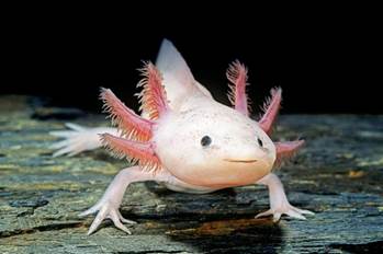
- The axolotl (Ambystoma mexicanum) is an aquatic salamander renowned for its ability to regenerate its spinal cord, heart and limbs. These amphibians also readily make new neurons throughout their lives.
- In 1964, researchers observed that adult axolotls could regenerate parts of their brains, even if a large section was completely removed. But one study found that axolotl brain regeneration has a limited ability to rebuild original tissue structure.
|
AXOLOTL
The axolotl is a paedomorphic salamander. Paedomorphosis is an alternative process to metamorphosis in which adults retain larval traits at the adult stage. It is frequent in newts and salamanders, where larvae reach sexual maturity without losing their gills. Axolotls are thus unusual among amphibians in that they reach adulthood without undergoing metamorphosis. Instead of taking to the land, adults remain aquatic and gilled. The species was originally found in several lakes underlying Mexico City, such as Lake Xochimilco and Lake Chalco.
They are listed as critically endangered in the wild, with a decreasing population of around 50 to 1,000 adult individuals, by the International Union for Conservation of Nature and Natural Resources (IUCN) and are listed under Appendix II of the Convention on International Trade in Endangered Species (CITES). Axolotls are used extensively in scientific research due to their ability to regenerate limbs, gills and parts of their eyes and brains. Axolotls were also sold as food in Mexican markets and were a staple in the Aztec diet.
|

Cell Functions
- Different cell types have different functions. They are able to specialize in certain roles because they each express different genes. Understanding what types of cells are in the brain and what they do helps clarify the overall picture of how the brain works. It also allows researchers to make comparisons across evolution and try to find biological trends across species.
- One way to understand which cells are expressing which genes is by using a technique called single-cell RNA sequencing (scRNA-seq). This tool allows researchers to count the number of active genes within each cell of a particular sample. This provides a “snapshot” of the activities each cell was doing when it was collected.
Single-cell RNA sequencing
- Single-cell RNA sequencing can provide information on the specific function of each cell in a sample.
- This tool has been instrumental in understanding the types of cells that exist in the brains of animals. Scientists have used scRNA-seq in fish, reptiles, mice and even humans. But one major piece of the brain evolution puzzle has been missing: amphibians.
|
SINGLE CELL SEQUENCING
Single-cell sequencing technologies refer to the sequencing of a single-cell genome or transcriptome, so as to obtain genomic, transcriptome or other multi-omics information to reveal cell population differences and cellular evolutionary relationships.
Single-cell RNA sequencing (scRNA-seq), for example, can reveal complex and rare cell populations, uncover regulatory relationships between genes, and track the trajectories of distinct cell lineages in development.
Traditional sequencing methods can only get the average of many cells, unable to analyze a small number of cells and lose cellular heterogeneity information.
|
Process of Cell regeneration: The mechanism explained
- Scientists focused on the telencephalon of the axolotl.
- In humans, the telencephalon is the largest division of the brain and contains a region called the neocortex, which plays a key role in animal behavior and cognition. Throughout recent evolution, the neocortex has massively grown in size compared with other brain regions. Similarly, the types of cells that make up the telencephalon overall have highly diversified and grown in complexity over time.
- Scientists used scRNA-seq to identify the different types of cells that make up the axolotl telencephalon, including different types of neurons and progenitor cells. Progenitor cells are cells that can divide into more of themselves or turn into other cell types.
- Scientists have identified what genes are active when progenitor cells become neurons, and found that many genes pass through an intermediate cell type called neuroblasts before becoming mature neurons. This was previously unknown to exist in axolotls but now scientists have found out that this intermediate cell type stage exists in Axolotls.
Brain regeneration in Axolotls happens in three main phases.
- The first phase starts with a rapid increase in the number of progenitor cells, and a small fraction of these cells activate a wound-healing process.
- In phase two, progenitor cells begin to differentiate into neuroblasts.
- Finally, in phase three, the neuroblasts differentiate into the same types of neurons that were originally lost.
- Scientists observed that the severed neuronal connections between the removed area and other areas of the brain get reconnected in Axolotls. This rewiring indicates that the regenerated area had also regained its original function.
https://www.downtoearth.org.in/blog/science-technology/axolotls-can-regenerate-their-brains-these-adorable-salamanders-are-helping-unlock-mysteries-of-brain-evolution-regeneration-84688


